


 The main cause of achalasia is degeneration of the nerve cells in the esophagus (the food pipe). The exact reason why this happens is not known. The loss of nerve cells in the esophagus causes two major problems that interfere with swallowing.
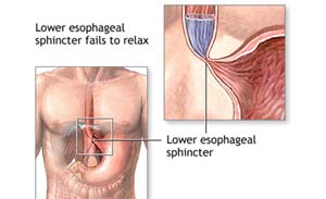
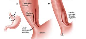
Firstly, the muscles that line the esophagus do not contract normally, so that swallowed food is not pushed forward through the esophagus and into the stomach properly. Secondly the lower esophageal sphincter (LES), a valve made of of muscles, does not relax with swallowing as it does in normal people. As a result, the esophagus above the persistently contracted LES starts to dilate, and large volumes of food and saliva can accumulate in the dilated esophagus.
The commonest symptom of achalasia is difficulty in swallowing. Patients get a sensation that swallowed food as well as liquids get stuck in the chest. This problem invariably progresses and becomes severe. Other symptoms include regurgitation of swallowed food and liquid, chest pain, heartburn, a sensation of fullness or a lump in the throat, hiccups, and weight loss.
The doctors can suspect achalasia based on the symptoms and order certain tests to confirm the diagnosis.
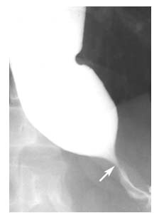
- Barium swallow: The barium swallow involves swallowing a milky liquid of barium while x-rays are taken. In patients with achalasia the barium swallow shows a narrow region at the lower end of the esophagus (bird’s beak) with a dilated esophagus above this
- Endoscopy: In this test the doctor passes a thin, lighted, flexible tube via the mouth to see the inside of the esophagus, LES, and stomach. Patients are usually given an intravenous sedative during the endoscopy procedure to make them sleepy and relaxed.
- Manometry: Manometry involves the passage of a thin tube through the mouth or nose into the esophagus. The tube is lined by numerous pressure sensors that convey pressures within the esophagus to a device. The tests measures the changes in pressures within the esophagus that are caused by the contraction of the muscles esophagus. Manometry is almost always used to confirm the diagnosis of achalasia. The test typically reveals three abnormalities in people with achalasia: high pressure in the LES at rest, failure of the LES to relax after swallowing, and an absence of useful (peristaltic) contractions in the lower esophagus.
The various options available for treating patients with achalasia do not stop or reverse the underlying loss of nerve cells in the esophagus or restore the normal peristaltic contractions. Rather, the treatments aim to weaken the lower esophageal sphincter (LES) muscle to the point that it no longer acts as a barrier to the passage of food and liquids.
Drugs: Two types of drugs, nitrates and calcium channel blockers, may be used for relaxing the LES muscle. However, in the long term the drug therapy is inconvenient, ineffective, and often associated with unpleasant side effects, such as headache and low blood pressure. Also, the drugs tend to become less effective when taken over a period of time.
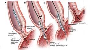
Balloon dilatation: This procedure is carried out under sedation. After preliminary endoscopy, a collapsed balloon is passed through the mouth and positioned in the region of the LES using guidance from an x-ray machine. It is then inflated in order to tear the muscle of the LES. The effect of a balloon dilatation in relieving the symptoms is temporary; most patients require multiple balloon dilatations at progressively shorter intervals and eventually require surgery. The most serious complication of a balloon dilatation is perforation (creation of a hole) of the esophagus, which occurs in 1% – 6% patients. The risk of a perforation increases with repeated dilatations. Because the LES has been made ineffective, 2% – 5% patients also develop gastroesophageal reflux disease (GERD) after balloon dilation. Also, after repeated dilatations the surgery, when it is eventually required, becomes more challenging. For these reasons, this treatment is best reserved for patients who are unfit for surgery due to old age or medical conditions which make administration of general anaesthetic risky. Young and fit patients not wishing to undergo surgery upon diagnosis of achalasia may be offered one dilatation, but are best counselled regarding surgery when the symptoms recur.
Botulinum toxin injection:
Injection of botulinum toxin (botox) into the LES under endoscopic guidance temporarily paralyzes the nerves that make the LES contract, thereby helping to relieve the obstruction. The relief of symptoms with this expensive therapy is short term (3 – 12 months). Also, repeated injections create scarring in the muscles of the LES, and make the subsequent surgery more challenging. This treatment, once quite popular, has fallen out of vogue recently.
Per Oral Endoscopic Myotomy (POEM)): This is a newly developed endoscopic procedure for treatment of achalasia cardia. The procedure is performed under general anaesthesia. An endoscope is passed into the esophagus and an incision is made in the lining (mucosa) of the esophagus. A tunnel is created below the mucosa and is dilated using a balloon. The muscle layers of the esophagus are then divided using an endoscopic knife. Finally the opening in the esophageal mucosa is closed. The attractiveness of the procedure lies in the fact that a surgery is avoided; however the procedure is not without significant drawbacks.The shortcomings of the POEM procedure are:
I. As this technique for treating achalasia has been around for only around three years, the long-term results in terms of relief from difficulty in swallowing remain unknown.
II. The procedure seems to be associated with complications (mostly minor) in a significant number of patients. Over 50% patients develop subcutaneous emphysema (swelling of arms, legs, face due to air in the layer under the skin). This happens as once an incision is made in the inner lining of the esophagus, the air used for keeping the esophagus open during the endoscopic procedure leaks out into the chest and can spread to the whole body and face. This air usually gets absorbed within a few days and does not require active treatment. Also, around 25% patients develop pneumothorax (air in the pleural cavity) and some of these patients are likely to require placement of a tube in the chest for removal of the air. Furthermore, nearly half the patients develop either fluid in the chest (pleural effusion) or inflammation of the lungs. It is important to note that risk of any of these complications in patients undergoing a laparoscopic cardiomyotomy is less than 2%.
Ren Z, Zhong Y, Zhou P, Xu M, Cai M, Li L, Shi Q, Yao L. Perioperative management and treatment for complications during and after peroral endoscopic myotomy (POEM) for esophageal achalasia (EA) (data from 119 cases). Surg Endosc. 2012 May 19. Read the abstract here.
I. The POEM procedure is aimed at dividing the LES that fails to relax (the underlying cause of achalasia). Effective and complete division of the LES (necessary for symptomatic relief) makes the patients prone to developing gastroesophageal reflux (tendency for the acid from the stomach coming back into the esophagus). Such a reflux is likely to develop in a fair number of patients after the POEM procedure. The POEM procedure does not address the issue of gastroesophageal reflux. On the other hand, surgeons performing laparoscopic cardiomyotomy, being aware of the likelihood of such reflux, always add a fundoplication to the procedure to prevent this problem (see below).
The POEM procedure, with its reported high incidence of immediate postoperative complications (albeit minor), propensity to produce undesirable effect of reflux and as yet unknown long-term results, is unlikely to replace the time-tested technique of laparoscopic cardiomyotomy in the near future.
The surgery performed to treat patients with achalasia is called cardiomyotomy (division of the muscles of the LES and upper end of stomach). In the past this surgery was performed through a large open incision in the upper abdomen. Today, it is carried out laparoscopically.
This operation is performed under general anaesthesia. The surgeon makes a small (1cm) incision in the upper abdomen and introduces a cannula or a tube inside the abdomen. He then inserts a telescope attached to a miniature video camera through the cannula that gives him and the operating team a magnified view of your internal organs on a video monitor. He will then place four other additional cannulas through tiny (5mm) cuts to accommodate special long instruments. At the surgery, the LES and the muscle layer in the upper part of the stomach are divided precisely under the magnified view. As division of LES makes the patient prone to gastro-esophageal reflux, the upper part of the stomach called fundus is rotated around and fixed in such a way that it creates a fundoplication or a valve. This prevents the acid from the stomach from coming back into the esophagus.
The patient does experience some amount of pain for about 12 to 24 hours after laparoscopic cardiomyotomy depending on individual tolerance. Also, some nausea and vomiting is not uncommon in the first 12 hours. Patients are always given medications to relieve the pain and take care of the nausea. Usually, the patient is allowed to drink fluids within 6 to 8 hours of surgery and is allowed soft blenderized food from the day after surgery. Activity is dependent on how the patient feels, but all patients are encouraged to get up and walk as soon as they are comfortable. Most patients go home within a 48 – 72 hours after the surgery. In general, patients recover completely within 10 – 15 days. All patients having a cardiomyotomy need to follow a blenderized diet for around 6 weeks as the area of surgery is healing. Also they are advised to avoid eat slowly, eat small frequent meals and avoid carbonated drinks. After the initial period of 6 weeks a patient is allowed to eat normal food.
In our society patients often prefer to take things easy for weeks after any operation because of a fear that they may harm themselves by being active. After laparoscopic cholecystectomy the recovery is quite rapid. Soon after returning home the patients are allowed all activities they feel comfortable with. Depending on the nature of their job, most patients are able to return to work within ten to fifteen days following a laparoscopic cardiomyotomy. Patients with light, desk jobs usually return in a few days while those involved in heavy lifting may require a little more time.
- Less pain from the incisions after surgery
- Shorter hospital stay
- Shorter recovery time
- Faster return to normal diet
- Faster return to work or normal activity
- Better cosmetic healing