




What are lymph nodes?
Lymph nodes are small, bean-shaped glands found throughout the body. They are part of the lymphatic system, which carries lymph fluid, nutrients, and waste material between body tissues and the bloodstream. The lymphatic system plays an important role in the body’s immune defense by filtering bacteria, viruses, and other harmful substances.
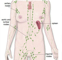
Where are the lymph nodes situated?
Lymph nodes are found throughout the body, either singly or in groups. They are commonly located in the neck, underarms, groin, abdomen, mesentery, and near major blood vessels such as the aorta and inferior vena cava.
What causes the lymph nodes to enlarge?
Lymph nodes usually enlarge because of infection, inflammation, or tumors. Swollen lymph nodes are generally painless and may develop near the affected area.
What does it mean when lymph nodes in two or more areas are enlarged?
Generalized enlargement of lymph nodes is known as generalized lymphadenopathy. Common causes include:
- – Viral infections
- – Bacterial infections including tuberculosis
- – Blood cancers such as leukemia, Hodgkin’s lymphoma, and Non-Hodgkin’s lymphoma
How are enlarged abdominal lymph nodes diagnosed?
Enlarged abdominal lymph nodes are usually detected through ultrasound or CT scan. Blood tests, endoscopy, and biopsy may also be required to determine the exact cause.
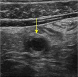
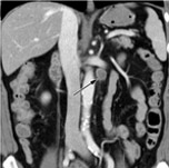
How is the lymph node biopsy performed?
A biopsy may be performed using FNAC, CT-guided biopsy, or surgical biopsy depending on the location of the enlarged lymph nodes. This helps establish an accurate diagnosis before treatment begins.
What if the diagnosis cannot be made after a CT-guided biopsy?
Sometime the sample obtained using a CT guided biopsy is too small for the pathologists to give a definite diagnosis of the cause of enlarged abdominal lymph nodes. Also, in some patients the enlarged lymph nodes may be located so close to important blood vessels or internal organs that it is risky to attempt a CT-guided biopsy.
Traditionally such patients required an open operation to obtain a sample from the lymph nodes. Today most abdominal lymph nodes that cannot be biopsied under CT guidance (which is the preferred method whenever possible, as it saves the patient from having a surgery) patients can have a laparoscopic biopsy of the enlarged lymph nodes.
How is laparoscopic lymph node biopsy performed?
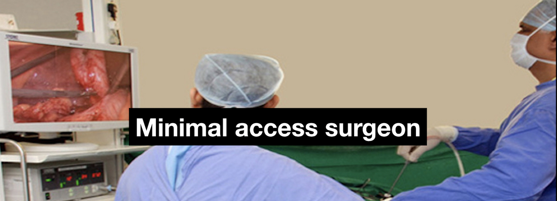
Laparoscopic lymph node biopsy is carried out under general anaethesia. The surgeon makes a small (about 1-cm) incision and places a short tube called a cannula through the abdominal wall. This cannula is connected to a special pump that pumps carbon dioxide gas. As the abdomen gets filled up with the gas, the abdominal wall is lifted up, thus providing the surgeon a space to work in. To look inside the abdomen, the surgeon passes a telescope connected to a miniature video camera through the cannula. The telescope that picks up the picture of the inside of the abdomen and transmits it to a television screen. The surgeon then exposes the area of enlarged lymph nodes and obtains a biopsy with the help of special, long instruments introduced inside the abdomen through two other cannulas and by observing the picture of the operative site on the television screen.
Usually, the sample is sent to a pathologist immediately for processing (called frozen section) when the patient is under anaesthesia. This helps the surgeon, as the pathologist can indicate whether the sample is adequate and is able to point to a tentative diagnosis. The final diagnosis often takes five to seven days as the sample must be processed in a particular manner and special tests carried out on it. In case the sample is deemed inadequate the surgeon can obtain more samples before the patient is brought out of anesthesia.
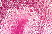
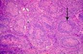
Images showing microscopic examination of a lymph node sample
Dr Bhandarkar’s publications and presentations related to laparoscopy for lymph nodal disorders
Publications and abstracts
1. Bhandarkar DS, Bhanushali P. Laparoscopic drainage of peripancreatic cold abscess. Surg Endosc Surg Endosc 2003; 17: 831.
2. Bhandarkar DS, Shah RS, Katara AN, Shankar M, Chandiramani VA, Udwadia TE. Laparoscopic biopsy in patients with abdominal lymphadenopathy. J Minim Access Surg 2007; 3:14-18.
Presentations, invited lectures & videos
1. Punjani RM, Bhandarkar DS. Diagnostic laparoscopy – a valuable tool for peeping inside the Pandora’s box. Annual Conference of Maharashtra Chapter of ASI, Pune, 2001.
2. Bhandarkar DS, Bhanushali P. Laparoscopic drainage of peripancreatic cold abscess. 5th Annual Conference of IAGES, Kolkata, 2002.
3. Bhandarkar DS, Bhanushali P. Laparoscopic drainage of peripancreatic cold abscess. Annual Conference of Maharashtra Chapter of ASI, Nasik, 2002.
4. Katara AN, Bhandarkar DS, Shah RS, Udwadia TE. Laparoscopy in management of abdominal lymphadenopathy. 6th National Conference of Endoscopic Surgery, Ludhiana, 2004.
5. Shankar M, Bhandarkar DS, Katara AN, Kochar R, Udwadia TE. Minimal access biopsy in patients with abdominal or thoracic lymphadenopathy. ELSA 2007, Hyderabad, 2007.
6. Bhandarkar DS, Behera RR, Katara AN, Udwadia TE. Laparoscopic biopsy in patients with abdominal lymph node enlargement. AMESCON 2012, Dubai, 2012.