


Rectal prolapse is a condition in which the rectum (the lower end of the colon, located just above the anus) becomes stretched and protrudes out of the anus. Weakness of the anal sphincter muscle is often associated with rectal prolapse at this stage, resulting in leakage of stool or mucus. While the condition occurs in both sexes, it is much more common in women than men.

Several factors may contribute to the development of rectal prolapse. It may come from a lifelong habit of straining to have bowel movements or as a late consequence of the childbirth process. Rarely, there may be a genetic predisposition. It seems to be a part of the aging process in many patients who experience stretching of the ligaments that support the rectum inside the pelvis as well as weakening of the anal sphincter muscle. Sometimes rectal prolapse results from generalized pelvic floor dysfunction, in association with urinary incontinence and pelvic organ prolapse as well. In most cases, however, no single cause is identified.
A doctor can often diagnose this condition with a careful history and a complete anorectal examination. To demonstrate the prolapse, patients may be asked to sit on a commode and “strain” as if having a bowel movement as this brings the prolapse in clear view and makes the diagnosis easy.
Rarely, a rectal prolapse may be “hidden” or internal, making the diagnosis more difficult. In this situation, an x-ray examination called a video-defecogram may be helpful. This examination, which takes x-ray pictures while the patient is having a bowel movement, can also assist the physician in determining whether surgery may be beneficial and which operation may be appropriate. Anorectal manometry or measurement of pressures in the anal canal and function of the sphincter muscles around may also be required in some patients. All patients require a flexible sigmoidoscopy or internal examination of the large bowel to exclude any other pathology.
Although constipation and straining may contribute to the development of rectal prolapse, simply correcting these problems may not improve the prolapse once it has developed. There are different operations for the treatment of rectal prolapse.
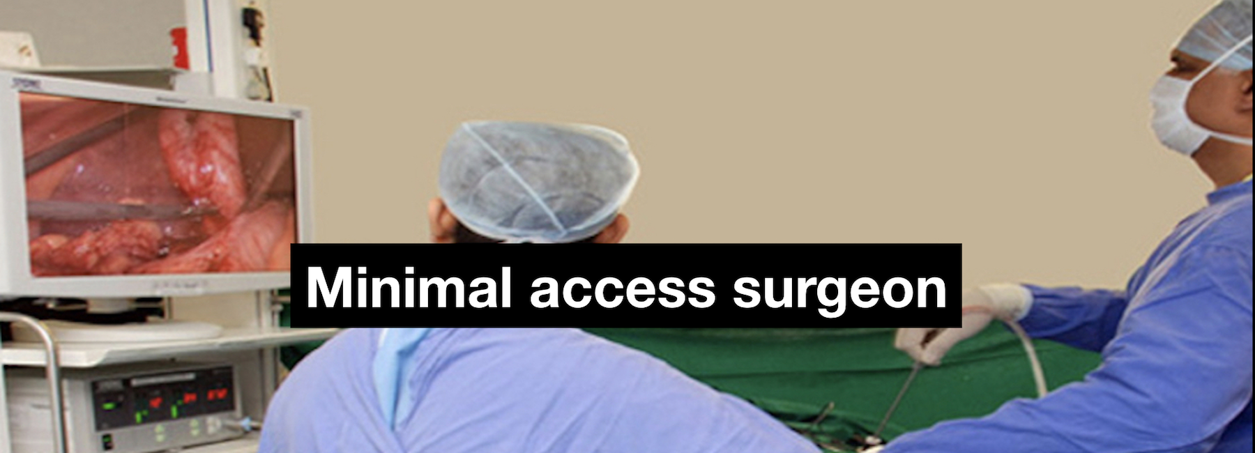
The operation to treat the rectal prolapse may be carried out from the abdomen or from rectal side. The surgeon takes into account factors such as age of the patient, physical condition, extent of prolapse and the results of various tests while deciding which route and which operation to use for treatment. By and large, patients who are fit to have a general anaesthesia benefit from a laparoscopic rectopexy. This operation is performed under general anaesthesia. The surgeon passes a cannula (a narrow tube) into the abdomen in the region of the umbilicus. A telescope connected to a camera is passed inside the abdomen through the cannula. The camera is connected to a television monitor so that a magnified view of the patient’s internal organs appears on the screen. The surgeon and his team conduct the operation by observing the television screen. The surgeon then inserts other cannulas and passes long, thin instruments inside the abdomen through them. Using these instruments the lowermost part of the large intestine – the rectum is freed up from its attachments. The rectum is then straightened up so that the prolapse gets pulled up. The rectum is then fixed to the bone behind called sacrum either with a few sutures or often by fixing a piece of mesh (net) between the bone and the rectum. This causes the rectum to stay fixed in the new, elevated position and prevents it from sliding down again.

The patient is allowed to have liquids within 6 – 8 hours of surgery and food the day after. The patient generally gets discharged within 48 – 72 hours of surgery and is prescribed a mild laxative for about a month so that he or she does not strain during bowel movement.
A great majority of patients are completely relieved of symptoms, or are significantly helped, by the appropriate procedure. Success depends on many factors, including the status of a patient’s anal sphincter muscle before surgery, whether the prolapse is internal or external, the overall condition of the patient. If the anal sphincter muscles have been weakened, either because of the rectal prolapse or for some other reason, they have the potential to regain strength after the rectal prolapse has been corrected. It may take up to a year to determine the ultimate impact of the surgery on bowel function. Chronic constipation and straining should be avoided after surgery.
What are the advantages of a laparoscopic rectopexy?
- Less pain from the smaller incisions after surgery
- Shorter hospital stay
- Shorter recovery time
- Faster return to normal diet
- Faster return to work or normal activity
- Better cosmetic healing (almost invisible scar)
Publications and abstracts
- Bhandarkar DS, Tamhane RG. Reduction of complete rectal prolapse. Trop Doct 1992; 22: 180.
- Bhandarkar DS. Delorme’s operation for complete rectal prolapse. Surgery 1999; 4:12-15.
- Bhandarkar DS, Katara AN. Laparoscopic mesh rectopexy: How I do it? J Colorectal Dis. 2007.
Book chapters
- Bhandarkar DS. Rectal prolapse: pathogenesis and clinical presentation. In, Clinical Book, 5th Instructional Course in Coloproctology, 2001
Presentations, invited lectures & videos
- Bhandarkar DS. Rectal prolapse: Etiopathogenesis and clinical presentation. Symposium on Rectal Prolapse at the Fifth Basic Instructional Course on Colorectal Surgery, Singhania Hospital, Thane, 2001.
- Bhandarkar DS. Delorme’s operation for rectal prolapse. Symposium on Rectal Prolapse at the Fifth Basic Instructional Course on Colorectal Surgery, Singhania Hospital, Thane, 2001.
- Pradhan C, Bhandarkar DS, Bhanushali HS. Laparoscopic rectopexy for complete procidentia. Annual Conference of Maharashtra Chapter of ASI, Thane, 2004.
- Bhandarkar DS, Pradhan C, Bhanushali HS. Laparoscopic rectopexy for rectal procidentia. 6th National Conference of Endoscopic Surgery, Ludhiana, 2004.
- Kochar R, Bhandarkar DS, Katara A, Udwadia TE. Laparoscopic mesh rectopexy for complete rectal prolapse. Annual Conference of Association of Colon Rectal Surgeons of India. Mumbai, 2008.
- Bhandarkar DS. Abdominal procedures for rectal prolapse. ASCRSI Postgradute Course, Thane, 2008.
- Bhandarkar DS. Laparoscopic rectopexy: master video. Annual Conference of Association of Colon & Rectal Surgeons of India, Goa, 2009.
- Bhandarkar DS. Laparoscopic rectopexy – How I do it? ASICON 2009, Coimbatore, 2009.
- Bhandarkar DS. Laparoscopic rectopexy – tricks for successful surgery. ASICON 2010, New Delhi, 2010.
- Bhandarkar DS. Ten steps to laparoscopic mesh rectopexy. ELSA2011, Singapore, 2011.
- Bhandarkar DS, Behera RR, Katara AN, Udwadia TE. Laparoscopic mesh rectopexy for full-thickness rectal prolapse. AMESCON 2012, Dubai, 2012.
- Bhandarkar DS. Rectal prolapse: abdominal versus perineal procedures. Which to do and when? Saifee Hospital Colorectal Surgery Update, Mumbai, 2013.