




Empyema
What is an empyema?
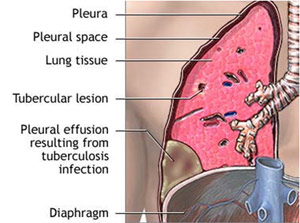
The pleural space is the space between the outer surface of the lung and the inside of the chest wall (rib cage). Normally, it contains a small amount of fluid that acts as a lubricant, allowing the lungs to move smoothly during breathing.
When a large amount of fluid collects in this space, it is called a pleural effusion. This fluid can become infected, providing an ideal environment for bacteria to grow, eventually turning into pus. This collection of pus in the pleural cavity is known as empyema.
How does an empyema develop?
Empyema usually develops after an infection in the nearby lung, such as pneumonia or tuberculosis.
The disease progresses through three stages:
- Exudative Phase – Fluid-like pus collects in the pleural cavity.
- Fibrino-purulent Phase – Fibrous bands (fibrin) develop, dividing the fluid into pockets.
- Organizing Phase – A thick fibrous peel forms around the lung, preventing it from expanding normally.
What symptoms does the patient experience?
Patients may experience:
- Persistent fever
- Cough
- Shortness of breath
- Chest pain
These symptoms often continue despite antibiotic treatment. When caused by tuberculosis, patients may also have weight loss, poor appetite, and prolonged illness.
How is an empyema diagnosed?
Diagnosis usually involves:
- Chest X-ray showing pleural effusion
- Pleural fluid aspiration using a fine needle
- Laboratory tests confirming pus or bacterial infection
- Ultrasound or CT scan to determine whether the fluid is free-flowing or trapped in pockets (loculated)

X-ray showing right-sided empyema |
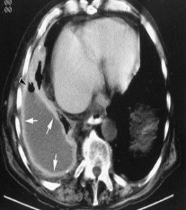
CT scan showing a loculated right-sided empyema |
How is an empyema treated?
Treatment follows three main principles:
- Appropriate antibiotics to control infection.
- Complete drainage of infected pleural fluid.
- Full expansion of the lung if trapped by fluid or fibrous tissue.
Early-stage empyema may be treated with needle aspiration or a chest tube. More advanced disease often requires thoracoscopic surgery to remove infected material and allow the lung to expand fully.
How is an empyema treated by thoracoscopic surgery?
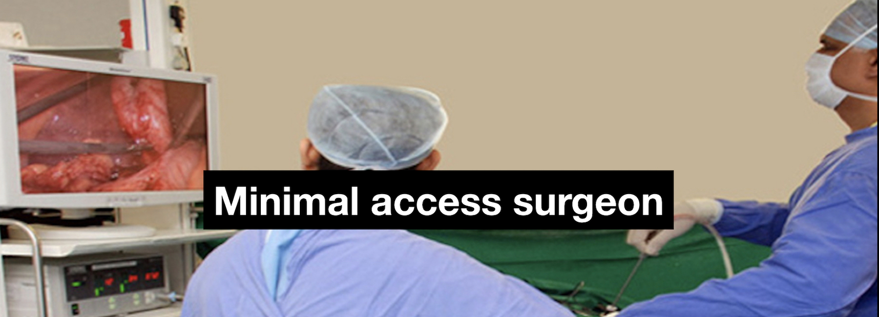
Thoracoscopic surgery is performed under general anesthesia through three or four small incisions. A camera (thoracoscope) and surgical instruments are inserted through these ports to remove infected fluid, debris, and fibrous tissue.
The surgeon ensures that the lung fully expands before completing the procedure. In 10–20% of advanced cases, conversion to an open surgery may be necessary.
What happens after the surgery?
A chest tube is placed after surgery to remove any remaining fluid and air. The tube is generally removed within 3–4 days, although some patients may require a longer hospital stay if drainage continues.
What are the advantages of thoracoscopic treatment?
- Less postoperative pain
- Shorter hospital stay
- Faster recovery
- Earlier return to work and normal activities
- Better cosmetic results due to smaller incisions
Resources
- Medscape – Empyema Overview
- Healthline – Empyema
- Medscape – Empyema Management
- CTSNet – Thoracoscopic Decortication