Empyema

The pleural space is the space between the outer surface of the lung and inside of the chest wall or rib cage. Normally, it contains a small amount of fluid that acts as a lubricant and facilitates movement of the lung with the diaphragm and chest wall. When a large quantity of fluid collects in the pleural space it is called a pleural effusion. A pleural effusion provides a rich culture medium for bacteria to grow in and may get converted to a collection of pus. Such a collection of pus in the pleural cavity is called empyema.
Empyema
An empyema usually develops following an episode of infection in the adjacent lung like pneumonia or tuberculosis. The formation of empyema can be divided into three phases: exudative, fibrino-purulent and organizing. During the first or exudative phase, pus (that is still fluid) accumulates in the pleural space. In the second or the fibrino-purulent phase multiple bands or septae of a material called fibrin start to develop within the pleural space. If the empyema is not treated at this stage, a thick membrane (called peel) forms and sticks to the lung. Once the peel is formed the lung is unable to expand completely or gets “trapped” and the empyema persists.
Patients with pneumonia often develop some fluid in the pleural space (pleural effusion) but this usually resolves. Patients with empyema have symptoms such as fever, cough, breathlessness and chest pain. These patients do not feel better even after a course of antibiotics, and in particular their fever persists. Patients who have an empyema secondary to tuberculosis are often ill for a long time and may have poor appetite and may have lost weight.
An x-ray of the chest usually shows a pleural effusion. When empyema is suspected a sample of the fluid is obtained with the help of a fine needle. The diagnosis of empyema is made when the pleural fluid reveals pus and when microscopic and microbiological tests show the fluid to contain bacteria or micro-organisms. An ultrasound scan or a CT scan is often required when the diagnosis of emypema is considered to check whether the fluid in the pleural space is free-flowing or has become loculated.
X-ray showing empyema on right side
CT scan showing a loculated right-sided empyema
The treatment of empyema involves three main principles:
  1. Use of appropriate antibiotics to control infection
  2. Complete evacuation of the infected pleural fluid
  3. Complete expansion of the lung if it is trapped because of the fluid or a fibrous peel
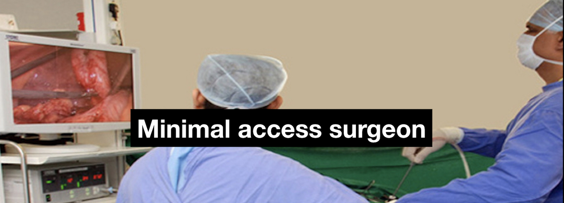
When the fluid is thin (in the early stages) it is possible to evacuate it completely by aspiration with the help of a needle or by placing a tube in the chest to drain it out. In the more advanced stage of the disease (as is often the case by the time the diagnosis is made) the thick fluid or the pus may not drain out through a tube. That is where thoracoscopic surgery helps in clearing all the fluid/ pus, clearing up all the loculi and allow the lung to expand.
The procedure is a major procedure and is undertaken under general anaesthesia. Three to four small incisions are made over the chest and tubes called ports are placed. A telescope is passed through one of the ports and instruments through the others. The aim of the surgery is to clear all the fluid, infected debris and the peel (if any) from the lung. At the end of the surgery complete expansion of the lung is confirmed. In late or difficult cases (10 – 20%) the procedure may have to be converted to an open operation through a long incision to satisfactorily complete the surgery. The later in the course of the disease the surgery is performed, the higher are the chances of the patient requiring an open operation.
After any surgery on the chest / lungs it is necessary to place a tube in the chest to remove the fluid and some air that may leak from the lung. This is kept in as long the fluid continues to drain. Most often the tube can be removed within three to four days but rarely, the leakage of fluid and air may continue for longer period of time and patient may require to stay in the hospital longer.
http://emedicine.medscape.com/article/298485-overview http://www.healthline.com/health/empyema http://emedicine.medscape.com/article/1970123-overview http://www.ctsnet.org/article/thoracoscopic-decortication
  1. Less pain from the incisions after surgery
  2. Possibly a shorter hospital stay than after an open operation. Often, how soon a patient is able to go home depends on the severity of the empyema and how soon the drainage tube placed in the chest stops draining.
  3. Shorter recovery time
  4. Faster return to work or normal activity
  5. Better cosmetic healing

Download Empyema Brochure