


Traditionally all operations on the chest have been performed by the open method that involves making a large incision – either a vertical midline cut splitting the breastbone (sternum) or one on the side and behind the chest through the rib cage. These large incisions provide excellent access to the organs within the chest but have their disadvantages. Large incisions cause significant postoperative pain and disability. The pain is likely to inhibit the patient’s ability to breathe deeply after surgery and clear the lungs of secretions. This may result some parts of the lung not opening up or inflating adequately (collapse or atelectasis). This often leads to fever after surgery and delays discharge from the hospital. Also the larger cuts are more prone to infection.

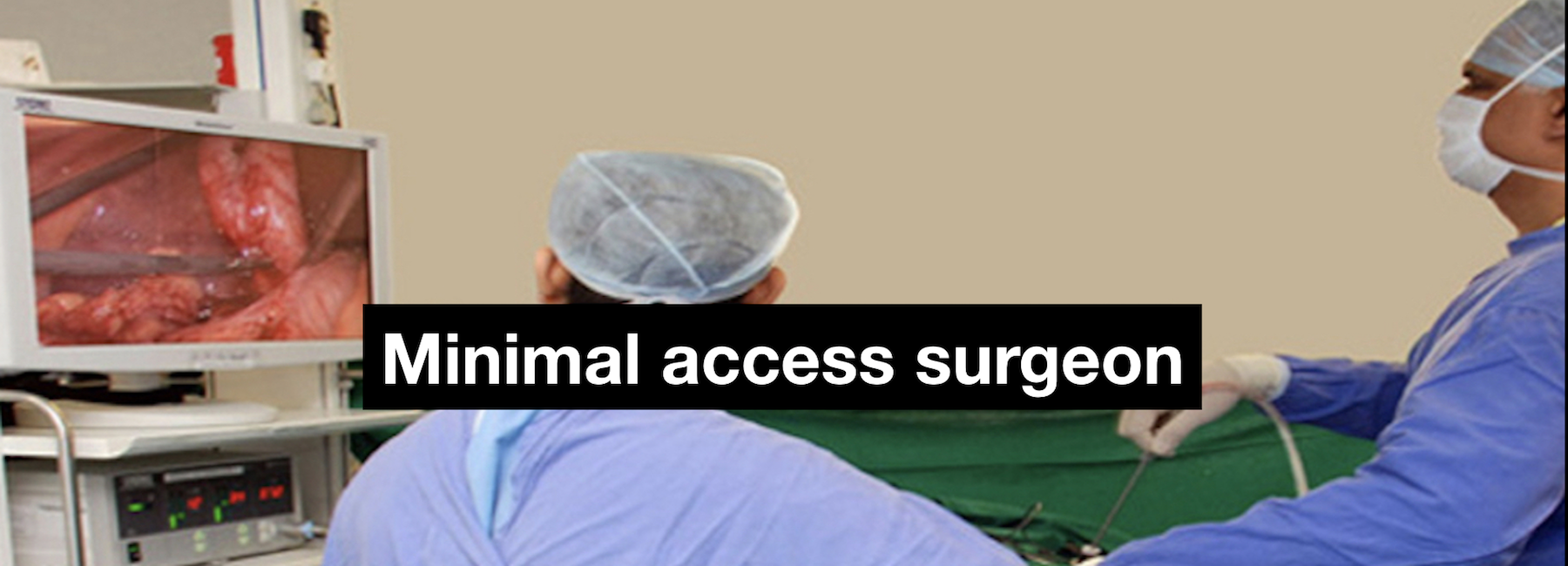
The operation is performed under general anaesthesia. During thoracoscopic operations the surgeon makes three to four small (about 1-cm) incisions and places tube called cannulas through the chest wall. To look inside the chest, the surgeon passes a telescope connected to a miniature video camera through one of the cannulas. The video camera picks up the picture of the inside of the chest cavity and transmits it to a television screen. The surgeon then carries out the operation with the help of special instruments introduced inside the chest through other cannulas and by observing the picture of the operative site on the television screen. At the end of the surgery usually one or two small tubes are placed in the chest cavity through the small incisions used for placing the cannulas. These tubes drain out the fluid or air that may collect inside after surgery. The tubes are removed after a few days.

Which operations can be performed using single incision (uniportal) thoracoscopic surgery?
Operations that can be performed using this technique include
- Sympathectomy
- Lung biopsy
- Pleural biopsy
- Blebecetomy or bullectomy for emphesematous bullae
Operations that can be performed using this technique include
- Sympathectomy
- Lung biopsy
- Pleural biopsy
- Blebecetomy or bullectomy for emphesematous bullae
You will wake up at the end of the operation. Some patients may require to be observed in an intensive care unit for a day or two after thoracoscopic surgery. You will be allowed to move about within a day or two of the surgery, although the tubes in the chest attached to bottles / bags need to be looked after during any activity. You will be given adequate painkillers to take care of the mild to moderate pain that occurs for the first couple of days. A very important part of the recovery after any thoracoscopic surgery is the breathing exercises that one needs to do with the help of a gadget called a spirometer and with the help of the physiotherapists. Both these activities ensure that the lung expands well and the risk of infection is minimised. The discharge after surgery depends on the disease for which the surgery has been carried out, e.g. patients having a thoracoscopic sympathectomy for sweaty palms can expect to go home the day after the surgery whereas someone having surgery for an empyema (collection of pus in the chest) may stay in the hospital for a week or even longer.
It is usually easier for patients to recover from a thoracoscopic operation as compared to an open chest surgery because the wounds from the incisions are much smaller. Sometime air leaks from the lung that don’t heal up quickly can keep you in the hospital a longer time and occasionally require additional treatment. A very small number of patients may require an additional procedure or placement of another tube in the postoperative period.
What are the advantages of thoracoscopic surgery?
- Less pain from the incisions after surgery
- Shorter hospital stay
- Shorter recovery time
- Faster return to work or normal activity
- Better cosmetic healing
- Less pain from the incisions after surgery
- Shorter hospital stay
- Shorter recovery time
- Faster return to work or normal activity
- Better cosmetic healing
http://my.clevelandclinic.org/services/heart/services/lungs-chest/video-assisted-thoracic-surgery
Publications and abstracts
- Katara AN, Samra SS, Bhandarkar DS. Thoracoscopic pericardial window for post-CABG pericardial effusion. Indian Heart J 2003; 55: 180-181.
- Shah R, Katara A, Bhandarkar DS. Thoracoscopic surgery: video assisted thoracic surgery (VATS) in children. Bombay Hospital J 2005; 47:350-53.
- Behera RR, Gouda B, Kulkarni A, Udwadia Z, Bhandarkar DS. Thoracoscopic and endovascular management of retained hemothorax associated with an intercostal artery pseudoaneurysm. Ind J Chest Dis. 2014;56:37-39.
Presentations, invited lectures & videos
- Bhandarkar DS, Katara AN, Samra S. Thoracoscopic pericardial window for post-CABG pericardial effusion. Annual Conference of International College of Surgeons, Chennai, 2003.
- Mittal G, Katara A, Bhandarkar DS, Udwadia TE. Promising results of bilateral endoscopic sympathectomy for palmar hyperhidrosis. ASICON 2010, New Delhi, 2010.
- Katara A, Mittal G, Bhandarkar DS, Udwadia TE. Overnight stay bilateral thoracoscopic sympathectomy. MASICON 2011, Mumbai, 2011.
- Bhandarkar DS. How would I perform VATS? 2nd National Pulmonary Update, Mumbai, 2008.
- Bhandarkar DS. Video assisted thoracic surgery – an overview. Seminar on Pleural Diseases, Vadodara, 2009.
- Bhandarkar DS. Video assisted thoracoscopic surgery. 13th FIAGES Course, Ahmedabad, 2011.
- Bhandarkar DS. Video assisted thoracic surgery. 18th FIAGES Course, Bhubaneswar, 2011.
- Bhandarkar DS. Video assisted thoracoscopic surgery. 20th FIAGES Course, Lucknow, 2011.
- Behera RR, Bhandarkar DS, Udwadia ZF, Kulkarni A, Katara A, Udwadia TE. Minimally invasive treatment of fibrothorax secondary to leaking posterior intercostal artery pseudoaneurysm. IAGES 2012, Ahmedabad, 2012.
- Behera RR, Bhandarkar DS, Udwadia ZF, Katara A, Udwadia TE. Uniportal thoracoscopic lung biopsy, resection of emphysematous bulla and pleurodesis. IAGES 2012, Ahmedabad, 2012.
- Bhandarkar DS, Katara AN, Behera RR. Thoracoscopic plication of diaphragmatic eventration in an adult. 20th Conference of European Conference of General Thoracic Surgery, Essen, 2012.
- Behera RR, Shah R, Bhandarkar D. Thoracoscopic excision of posterior mediastinal neuroblastoma in a one-year old infant. AMASICON 2012, Coimbatore, 2012.
- Bhandarkar DS. Video assisted thoracic surgery. 23rd FIAGES Course, Aurangabad, 2012.
- Bhandarkar DS. Video assisted thoracic surgery. 24th FIAGES Course, Patna, 2012.